Abstract
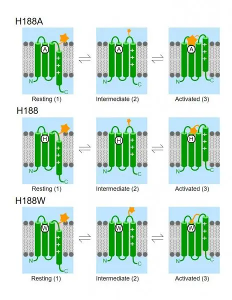
Voltage-clamp fluorometry (VCF) supplies information about the conformational changes of voltage-gated proteins. Changes in the fluorescence intensity of the dye (TAMRA) attached to a part of the protein that undergoes a conformational rearrangement upon the alteration of the membrane potential by electrodes constitute the signal. The VCF signal is generated by quenching and dequenching of the fluorescence as the dye traverses various local environments. Here we studied the VCF signal generation, using the Hv1 voltage-gated proton channel as a tool, which shares a similar voltage-sensor structure with voltage-gated ion channels but lacks an ion-conducting pore. Using mutagenesis and lipids added to the extracellular solution we found that the signal is generated by the combined effects of lipids (enhance fluorescence) during movement of the dye relative to the plane of the membrane and by quenching amino acids. Our 3-state model recapitulates the VCF signals of the various mutants and is compatible with the accepted model of two major voltage-sensor movements.
Link: https://www.nature.com/articles/s42003-022-04065-6
DOI: https://doi.org/10.1038/s42003-022-04065-6