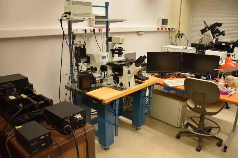
Name of the system: OLYMPUS FluoView FV1000 Confocal Microscope
Location of the instrument: Life Science Building 2nd floor; 2.045 Room
Organisation: Sándor Damjanovich Cell Analysis Core Facility, Department of Biophysics and Cell Biology, Faculty of Medicine, University of Debrecen
Core Facility Homepage: biophys.med.unideb.hu/dszl E-mail: dszl@med.unideb.hu
Contact: Dr. Mocsár Gábor (Tel.: 66566) E-mail: mocsgab@med.unideb.hu
Registration:
- Only authorized and trained Users are allowed to use the Equipment in the Core Facility!
- Users need to attend an instrument specific training program before being allowed to sign up to use the equipment independently.
- Users need to attend an instrument specific training program before being allowed to sign up to use the equipment independently.
Online instrument booking: link
Specifications: Product webpage
Olympus FluoView 1000 konfokális lézerpásztázó mikroszkóp és fluoreszcencia korrelációs spektroszkóp. 456, 470, 488, 514, 543 és 633 nm-es megvilágítás, 3 fluoreszcens és korrelációs detektor.
Műszer rövid ismertetése:
Mikroszkópos tomográfiára (optikai szeletelés), illetve molekuláris mobilitás és együttmozgás kimutatására alkalmas berendezés. A mikroszkóp 3 lézere 456, 470, 488, 514, 543 és 633 nm-es gerjesztési hullámhosszakat biztosít. A mintából egyidejűleg 3 fluoreszcencia – ebből 2 spektrális feloldású – és egy áteső fényű (DIC kontrasztozású) jel detektálható. Pásztázással 0,5 μm optikai szeletvastagságú képek készíthetők, melyekből rekonstruálható a minta 3D képe, a biomolekulák térbeli eloszlása, kolokalizációja. A mikroszkóphoz egyedi tervezésű, háromcsatornás fluoreszcencia korrelációs és keresztkorrelációs spektroszkóp (FCS, FCCS) kapcsolódik, mellyel a sejt kiválasztott (0,3 μm3-es) térfogatelemében végezhető FCS mérés.
Potential use:
- co-localization analyses with 200 nm resolution
- multichannel confocal imaging
- multichannel fluorescent correlation and cross-correlation spectroscopy
- optical sectioning, 3D reconstruction
- FRET, FRAP measurements
